


New cell atlas could unlock clues about disease
Interview with Professor Joe Ecker, Salk Institute for Biological Studies, San Diego, California
Estimated reading time: 9 minutes
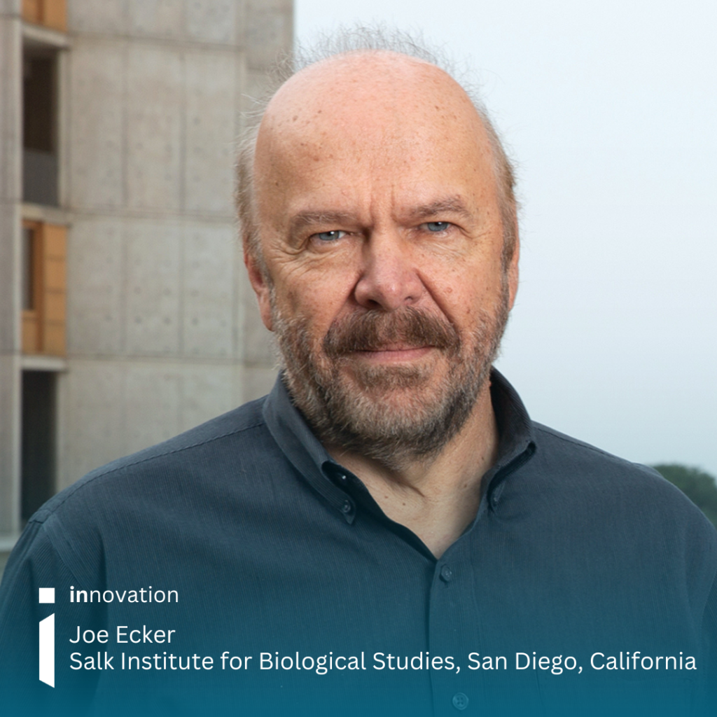

Researchers profiled 86,689 cell nuclei across 16 human tissue types, linking the switches that turn our genes on and off to disease risk. Professor Joe Ecker, of the Salk Institute for Biological Studies in San Diego, California, was involved in the research and shares the progress so far and his hopes for how the study might be further scaled
A new cell atlas is shedding light on how genetic risk contributes to disease and could ultimately help researchers better understand rare conditions. Scientists hope to expand the project, creating a resource that may help identify previously unexplained genetic causes of disease.
The single-cell atlas simultaneously mapped 3D genome architecture (how strings of DNA two metres long are folded inside a cell nucleus) and the chemical tags attached to DNA that modify it and can turn genes on and off (DNA methylation).
The team, which included researchers at the Salk Institute and the Arc Institute, analysed 86,689 human cell nuclei spanning 16 tissues, 35 major cell types and 206 subtypes. Their work gives a detailed view of how cells with identical DNA become vastly different cell types, and of which cells genetic risk factors are most likely to act in.
Researchers measured the 3D genome architecture and DNA methylation to compare what they say about a cell’s identity. Results showed that although the two pictures often agreed, sometimes they did not.
By superimposing the atlas with known disease-associated genetic variants, researchers identified specific cell types linked to conditions including atrial fibrillation, bipolar disorder, schizophrenia and blood-glucose regulation.
To make the findings accessible, the team created a free, interactive atlas, hosted by the Arc Institute, that allows scientists to explore how DNA is organised and regulated across the human body.
The work is part of the National Institutes of Health’s 4D Nucleome (NIH 4DN) program1, which aims to understand how the genome is organised in space and time to regulate gene expression in health and disease. It was published alongside five other papers from the programme in Science and another three in Science Advances.
What the atlas reveals
Professor Joe Ecker, who sits as the Salk International Council Chair in Genetics, was one of the co-corresponding authors of the research.
As well as DNA methylation the cell atlas also reports on DNA folding and modification, which help control when genes are switched on and off. Joe explains how they had looked at the non-coding parts of the genome—the areas that don’t make a protein. These regions are important in controlling the expression of that gene.
He explains that identifying which genetic variations are responsible for causing disease remains challenging because every person’s genome contains differences, but the atlas helps with this. The maps they are making identify the regions in the genome that are likely to be involved in switching genes on and off.
“It is a very cell-type-specific map where we have identified over 200 cell types, and each cell type has different switches. If you have a part of the gene you suspect might be involved in the disease, this helps you to narrow down where that change might be.”
Because most common disease risk variants are found in these control regions rather than in the genes themselves, the map helps researchers connect a genetic risk to specific cells and organs.
For instance, Joe explains that if someone is studying schizophrenia, they could use the map to see which switches are active in the specific brain cells linked to the condition.
Understanding disease
Speaking of how the atlas can help with the understanding of disease, particularly in the rare disease sector, Joe says: “In about half the individuals where their genome is sequenced, you can identify a causal event contributing to that rare disease. The other half you cannot, and most of those events and changes in the DNA are likely outside, very far outside, of the genes.
“What we are doing here is mapping all of the regions in these different body tissues where you likely have switches that control the genes, turning them on and off.”
Joe explains as around half of rare diseases can’t be easily identified by looking within the protein coding part of the genes, the atlas gives a “spotlight” to look in other regions of the genome that have chemical tags, to see if there’s any genetic changes, deletions or duplications.
He says: “These maps, or particularly the DNA methylation, have much more information in the genome than other kinds of technologies, which allow you to look at these switches.”
Many rare diseases are first detected during a child’s development. Joe said an area where there is still a gap in understanding is early-stage development, when changes that may contribute to rare conditions could occur.
Alongside insights into disease the study also revealed unexpected differences in how genes are regulated. The difference between what the 3D genome and DNA methylation showed was a surprising outcome.
“We were looking at two different ways to regulate gene expression. One has the folding, and one is the chemical events. What we found was that sometimes the information about what the cell was doing was in the folding part of the analysis, and other times it was in the chemical tags analysis. So, there is more than one way to regulate a gene.”
Likewise, another discovery came to light during a companion study that Joe’s lab contributed to2. For years, scientists had believed microglia—the brain’s immune cells—persist from embryonic development throughout our lives. However, the researchers found that between the ages of 50 and 75, these cells are progressively replaced by a new microglial population whose epigenetic profile points to an origin in monocytes, immune cells from the blood.
In donors over 79, almost all hippocampal microglia were of the new type. Because these replacement cells still look and behave like microglia, the shift is invisible to standard gene-expression tests; the researchers detected it through DNA methylation, which preserves the chemical marks left over from a cell’s earlier life.
Atlas could aid AI models for predictive medicine
The atlas was a five-year project, and they had to develop the right technology before even starting the work. Joe explains: “First we had to develop the technology to be able to identify these chemical tags and the folding, and we did that simultaneously.”
He adds: “We have been applying it to the human body tissues. With recent focus on the brain. But this is just the beginning. We have looked at a dozen-plus tissues and identified a few hundred cell types, but there are many other tissues to be examined, and we are hoping to continue to do that.”
AI-driven discovery is a massive area of development now, and the atlas could help accelerate this. Before they had even published the paper about the study, the team had been contacted by AI companies who were working to build models for predictive medicine.
Joe says the firms want to understand how genetic changes match epigenetic data (changes in gene activity that don’t alter the DNA code).
He adds: “Our map here is one of the largest and encompasses many tissues in the body. We put the data out there for those groups to use to build AI models.”
The data are freely available through open-access platforms including the Gene Expression Omnibus, Hugging Face, GitHub and Zenodo.

Hopes for the future
During the research, one challenge was the availability of tissue to analyse, and Joe says they are thankful to the families, particularly those of children and adolescents, who agreed to donate.
“We really hope that families consider these donations. It is super important for moving science forward. It has been somewhat of a limitation to our research. We have funding, we have the technology, and what we are lacking in is donor tissues. I really want to make a plea for folks to consider that.”
If an individual hasn’t registered as a donor prior to their death, consent for tissue donation can still be provided by their next of kin (in the US and other opt-in donation systems). This allows tissue to be used not only for transplantation but also for research studies.
Joe adds, “We really desperately would like people to consider donation, especially in children and adolescents, so we can begin to make maps to help other families where the diseases are really not understood, because the switches change over developmental time. If we make an atlas of adults, as we’ve done here, it provides some information about what the end stage is. But we really need to have these maps, atlases of the entire profile of human development.”
The atlas is available online athumancellepigenomeatlas.arcinstitute.org3and going forward Joe hopes they can expand the study.
He shares: “The challenge is to make the maps more comprehensive, so they can be used for all kinds of diseases. We need to scale up the study, so we encompass more developmental stages, more tissues.”
Connect with Joe
References
[1] https://commonfund.nih.gov/4DNucleome
[2] Zemke, N. R. et al. Epigenetic and 3D genome reprogramming during the aging of the human hippocampus. Science 393, eadt8307 (2026). https://doi.org/10.1126/science.adt8307
[3] https://humancellepigenomeatlas.arcinstitute.org/
innovation spotlights the future of RARE through a technological lens. To access more innovation articles click below.